Just What Is an RLU (Relative Light Unit)?

This post was contributed by guest blogger, Scott Messenger, Technical Support Scientist 2 at Promega Corporation.

It’s always an exciting time in the lab when you find a new assay to answer an important research question. Once you get your hands on the assay, it is always good to confirm it will work for your experimental setup. Repeating the control experiment shown in the technical manual is a great way to test the assay in your hands.

After running that first experiment of your assay, it looks pretty good. The trends of control and treatment are consistent. Time to get on with the experiments…but wait—the RLUs (Relative Light Units) are two orders of magnitude lower than the example data! I can’t show this data to my colleagues; it doesn’t match. What did I do wrong?

Relative Light Units, Measuring, Luminescence

This is a concern that we in Technical Services hear frequently. The concern is real, and I had this same thought when doing some of my first experiments using luminescence. When a question like this comes in, a Technical Service Scientist will make sure the experiment was performed as we described, and in most cases it is. We then start talking about RLUs (Relative Light Units).

Continue reading “Just What Is an RLU (Relative Light Unit)?”

New Uses for Old Drugs: Remdesivir and COVID-19

With the COVID-19 pandemic far from over in the United States and worldwide, the battle against the disease continues to intensify. Much hope has been pinned on vaccine development. However, vaccines are a long-term, preventative strategy. The immediate need for drugs to fight COVID-19 has accelerated efforts for a variety of potential treatments (see The Race to Develop New Therapeutics Against Coronaviruses).

The Remdesivir Origin Story

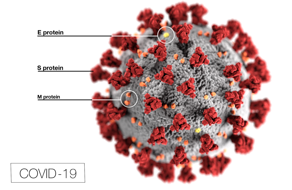
3d model of coronavirus

One drug that has received widespread attention is remdesivir. It was developed from research by Gilead Sciences that began in 2009, originally targeting hepatitis C virus (HCV) and respiratory syncytial virus (RSV) (1). At present, remdesivir is classified as an investigational new drug (IND) and has not been approved for therapeutic use anywhere in the world.

Continue reading “New Uses for Old Drugs: Remdesivir and COVID-19”

Choices for Measuring Luciferase-Tagged Reporter Pseudotyped Viral Particles in Coronavirus Research

Coronavirus (CoV) researchers are working quickly to understand the entry of SARS-CoV-2 into cells. The Spike or S proteins on the surface of a CoV is trimer. The monomer is composed of an S1 and S2 domain. The division of S1 and S2 happens in the virus producing cell through a furin cleavage site between the two domains. The trimer binds to cell surface proteins. In the case of the SARS-CoV, the receptor is angiotensin converting enzyme 2. (ACE2). The MERS-CoV utilizes the cell-surface dipeptidyl peptidase IV protein. SARS-CoV-2 uses ACE2 as well. Internalized S protein goes though a second cleavage by a host cell protease, near the S1/S2 cleavage site called S2′, which leads to a drastic change in conformation thought to facilitate membrane fusion and entry of the virus into the cell (1).  

CDC / Alissa Eckert, MS; Dan Higgins, MAMS

Rather than work directly with the virus, researchers have chosen to make pseudotyped viral particles. Pseudotyped viral particles contain the envelope proteins of a well-known parent virus (e.g., vesicular stomatitis virus) with the native host cell binding protein (e.g., glycoprotein G) exchanged for the host cell binding protein (S protein) of the virus under investigation. The pseudotyped viral particle typically carries a reporter plasmid, most commonly firefly luciferase (FLuc), with the necessary genetic elements to be packaged in the particle. 

To create the pseudotyped viral particle, plasmids or RNA alone are transfected into cells and the pseudotyped viruses work their way through the endoplasmic reticulum and golgi to bud from the cells into the culture medium. The pseudoviruses are used to study the process of viral entry via the exchanged protein from the virus of interest. Entry is monitored through assay of the reporter. The reporter could be a luciferase or a fluorescent protein.   

Continue reading “Choices for Measuring Luciferase-Tagged Reporter Pseudotyped Viral Particles in Coronavirus Research”

Designing a Reporter Construct for Analyzing Gene Regulation

Bioluminescent reporter assays are an excellent choice for analyzing gene regulation because they provide higher sensitivity, wider dynamic range and better signal-to-background ratios compared to colorimetric or fluorescent assays. In a typical genetic reporter assay, cells are transfected with a vector that contains the sequence of interest cloned upstream of a reporter gene, and the reporter activity is used to determine how the target sequence influences gene expression under experimental conditions. A second control reporter encoded on the same or a different plasmid is an essential internal control. The secondary reporter is used to normalize the data and compensate for variability caused by differences in cell number, lysis efficiency, cell viability, transfection efficiency, temperature, and measurement time. 

Basic Introduction to the Strategy of Reporter Gene Assays

For genetic reporter assays, using a secondary control vector with a weak promoter like PGK or TK to ensures that the control does not interfere with activation of your primary reporter vector. Transfection of high amounts of the control plasmid or putting the control reporter under control of a strong promoter like CMV or SV40 often leads to transcriptional squelching or other interference with the experimental promoter (i.e., trans effects). Reporter assays can also be used to quantitatively evaluate microRNA activity by inserting miRNA target sites downstream or 3´ of the reporter gene. For example, the pmirGLO Dual-Luciferase miRNA Target Expression Vector is based on dual-luciferase technology, with firefly luciferase as the primary reporter to monitor mRNA regulation and Renilla luciferase as a control reporter for normalization.

Here in Technical Services we often talk with researchers who are just starting their project and looking for advice on designing their genetic reporter vector. They have questions like:

  • How much of the upstream promoter region should be included in the vector?
  • How many copies of a response element will be needed to provide a good response?
  • Does the location of the element or surrounding sequence alter gene regulation?
Continue reading “Designing a Reporter Construct for Analyzing Gene Regulation”

Tips for Successful Dual-Reporter Assays

Updated 02/12/2021

Previously, we described some of the advantages of using dual-reporter assays (such as the Dual-Luciferase®, Dual-Glo® Luciferase and the Nano-Glo® Dual-Luciferase® Systems). Another post describes how to choose the best dual-reporter assay for your experiments. For an overview of luciferase-based reporter gene assays, see this short video:

These assays are relatively easy to understand in principle. Use a primary and secondary reporter vector transiently transfected into your favorite mammalian cell line. The primary reporter is commonly used as a marker for a gene, promoter, or response element of interest. The secondary reporter drives a steady level of expression of a different marker. We can use that second marker to normalize the changes in expression of the primary under the assumption that the secondary marker is unaffected by what is being experimentally manipulated.

While there are many advantages to dual-reporter assays, they require careful planning to avoid common pitfalls. Here’s what you can do to avoid repeating some of the common mistakes we see with new users:

Continue reading “Tips for Successful Dual-Reporter Assays”

I Have My Luciferase Vector, Now What?

Choosing and Optimizing Transfection Methods

Here in Technical Services we often talk with researchers at the beginning of their project about how to carefully design and get started with their experiments. It is exciting when you have selected the Luciferase Reporter Vector(s) that will best suit your needs; you are going to make luminescent cells! But, how do you pick the best way to get the vector into your cells to express the reporter? What transfection reagent/method will work best for your cell type and experiment? Do you want to do transient (short-term) transfections, or are you going to establish a stable cell line?

Continue reading “I Have My Luciferase Vector, Now What?”

Why You Don’t Need to Select a Wavelength for a Luciferase Assay

Promega kit depicted; test involves wavelength for a luciferase assay.

It’s a question I’m asked probably once a week. “What wavelength do I select on my luminometer when performing a luciferase assay?” The question is a good and not altogether unexpected one, especially for those new to bioluminescent assays. The answer is that in most cases, you don’t and in fact shouldn’t select a wavelength (the exception to this rule is if you’re measuring light emitted in two simultaneous luciferase reactions). To understand why requires a bit of an explanation of absorbance, fluorescence, and luminescence assays, and the differences among them.

Absorbance, fluorescence, and luminescence assays are all means to quantify something of interest, be that a genetic reporter, cell viability, cytotoxicity, apoptosis, or other markers. In principle, they are all similar. For example, a genetic reporter assay is an indicator of gene expression. The promoter of a gene of interest can be cloned upstream of a reporter such as β-galactosidase, GFP, or firefly luciferase. The amount of each of these reporters that is transcribed into mRNA and translated into protein by the cell is indicative of the endogenous expression of the gene of interest.

Continue reading “Why You Don’t Need to Select a Wavelength for a Luciferase Assay”

Eight Considerations for Getting the Best Data from Your Luminescent Assays

The stage is set. You’ve spent days setting up this experiment. Your bench is spotless. All the materials you need to finally collect data are laid neatly before you. You fetch your cells from the incubator, add your detection reagents, and carefully slide the assay plate into the luminometer. It whirs and buzzes, and data begin to appear on the computer screen. But wait!

Bad data
These data are garbage!

Don’t let this dramatic person be you. Here are 8 tips from us on things to watch out for before you start your next luminescent assay. Make sure you’ll be getting good data before wasting precious sample!

Continue reading “Eight Considerations for Getting the Best Data from Your Luminescent Assays”

Dual-Luciferase or Dual-Glo Luciferase Assay System? Which one should I choose for my reporter assays?

Confused woman

I’ve got a set of experiments planned that, if all goes well, will provide me with the answer I have been seeking for months. Plus, my supervisor is eagerly awaiting the results because she needs the data for a grant application, so I don’t want to mess it up. However, I am faced with a choice for my firefly and Renilla luciferase reporter assays: Do I use the Dual-Luciferase® Reporter Assay System or Dual-Glo® Luciferase Assay System? What’s the difference? How do I decide which to use? I’m so confused! Help!

Sound familiar? Not to worry! The choice is not difficult once you know how these assays work and how they differ.

Continue reading “Dual-Luciferase or Dual-Glo Luciferase Assay System? Which one should I choose for my reporter assays?”

Nano, Nano: Tiny Lipid Particles with Big Therapeutic Potential

cell-transfection-viafect-luciferase-assay

Getting DNA or RNA into cells can be a tricky business, and a variety of transfection reagents have been developed over the years to make the process easier. Lipid-based reagents are especially popular because they combine efficient transfection with relatively low toxicity.

When it comes to transfection, it pays to think small. Human cells range in volume from 20–40 µm3 (sperm cells) to as large as 4 million µm3 (mature egg cells, or oocytes). For several decades, transfection reagents have targeted this size range. However, breakthrough research involves leaving the “micro” realm and entering a world that was once the domain only of science fiction: nanotechnology.

Continue reading “Nano, Nano: Tiny Lipid Particles with Big Therapeutic Potential”