What’s In YOUR Protein? Optimizing Protease Digestions to Get the Inside Scoop

It’s time to analyze your protein and you are trying to decide where to begin. You are asking questions like: Which protease do I choose? How much enzyme should I use in my digest? How long should I perform my digest?

Unfortunately, there is no one-size fits all answer to this type of question other than… “well it depends.” All protease digests will be a balance between denaturing the protein sample to allow access to cleavage sites, optimizing conditions for the protease to function, and compatibility with your workflow and downstream applications. We provide general guidelines that work for most samples, but frequently you will need to optimize the conditions need for your specific sample and application.

Here, I use the example of a trypsin digest for downstream mass spectrometry to highlight key questions to ask and factors that can be optimized for any digest.

Continue reading “What’s In YOUR Protein? Optimizing Protease Digestions to Get the Inside Scoop”

New Recombinant Asp-N Mass Spec Protease: Improved Format and Reduced Price

Asp-N is a endoproteinase hydrolyzes peptide bonds on the N-terminal side of aspartic residues. The native form is isolated from Pseudomonas fragi. The majority of vendors currently provide a commercial product that consists of 2µg of lyophilized material in a flat bottom vial, and sold for $175–200 US. Formatting such a small amount of material in flat bottom vial can lead to inconsistent resuspension of the protease. Inconsistent working concentrations will lead to non-reproducible data. The current high price also prohibits large-scale use.

Are you looking for proteases to use in your research?
Explore our portfolio of proteases today.

The new recombinant Asp-N protease is cloned from Stenotrophomonas maltophilia and expressed in E. coli. Recombinant Asp-N has similar amino acid cleavage specificity as compared to native Asp-N. Digestion of a yeast extract with native and recombinant Asp-N produces very similar results. Providing 10µg lyophilized material in V-shaped vial with a visible cake enables more consistent re-suspension resulting in reproducible data. Due to improved yields the list price is now approximately 40% less when compared to native enzyme.
Learn more about this new recombinant Asp-N protease.

Mass Spec Analysis of PTMs Using Minimal Sample Material

DNA is organized by protein:DNA complexes called nucleosomes in eukaryotes. Nucleosomes are composed of 147 base pairs of DNA wrapped around a histone octamer containing two copies of each core histone protein. Histone proteins play significant roles in many nuclear processes including transcription, DNA damage repair and heterochromatin formation. Histone proteins are extensively and dynamically post-translationally modified, and these post-translational modifications (PTMs) are thought to comprise a specific combinatorial PTM profile of a histone that dictates its specific function.  Abnormal regulations of PTM may lead to developmental disorders and disease development such as cancer.

Continue reading “Mass Spec Analysis of PTMs Using Minimal Sample Material”

Optimized Detection of EPO-Fc in Human Biological Fluids

Recombinant erythropoietin (rhEPO) is often used as “doping agent” by athletes in endurance sports to increase blood oxygen capacity. Some strategies improve the pharmacological properties of erythropoietin (EPO) through the genetic and chemical modification of the native EPO protein. The EPO-Fcs are fusion proteins composed of monomeric or dimeric recombinant EPO and the dimeric Fc region of human IgG molecules. The Fc region includes the hinge region and the CH2 and CH3 domains. Recombinant human EPOs (rhEPO) fused to the IgG Fc domain demonstrate a prolonged half-life and enhanced erythropoietic activity in vivo compared with native or rhEPO.

Drug-testing agencies will need to obtain primary structure information and develop a reliable analytical method for the determination of EPO-Fc abuse in sport. The possibility of EPO-Fc detection using nanohigh-performance liquid chromatography−tandem mass spectrometry (HPLC−MS/MS) was already demonstrated (1). However, the prototyping peptides derived from EPO and IgG are not selective enough because both free proteins are naturally presented in human serum. In a recent publication, researchers describe the effort to identify peptides covering unknown fusion breakpoints (later referred to as “spacer” peptides; 2). The identification of “spacer” peptides will allow the confirmation of the presence of exogenous EPO-Fc in human biological fluids.

Are you looking for proteases to use in your research?
Explore our portfolio of proteases today.

A bottom-up approach and the intact molecular weight measurement of deglycosylated protein and its IdeS proteolytic fractions was used to determine the amino acid sequence of EPO-Fc. Using multiple proteases, peptides covering unknown fusion breakpoints (spacer peptides) were identified.

Results indicated that “spacer peptides” could be used in the determination of EPO-Fc fusion proteins in biological samples using common LC−tandem MS methods.

References

  1. Reichel, C. et al. (2012) Detection of EPO-Fc fusion protein in human blood: screening and confirmation protocols for sports drug testing.
    Drug Test. Anal. 4, 818−29.
  2. Mesonzhnik, N. et al. (2017) Characterization and Detection of Erythropoietin Fc Fusion Proteins Using Liquid Chromatography−Mass Spectrometry.
    J. of Proteome Res. 17, 689-97.

Analysis of a biosimilar mAb using Mass Spectrometry

Several pharmaceutical companies have biosimilar versions of therapeutic mAbs in development. Biosimilars can promise significant cost savings for patients, but the unavoidable differences
between the original and thencopycat biologic raise questions regarding product interchangeability. Both innovator mAbs and biosimilars are heterogeneous populations of variants characterized by differences in glycosylation,oxidation, deamidation, glycation, and aggregation state. Their heterogeneity could potentially affect target protein binding through the F´ab domain, receptor binding through the Fc domain, and protein aggregation.

As more biosimilar mAbs gain regulatory approval, having clear framework for a rapid characterization of innovator and biosimilar products to identify clinically relevant differences is important. A recent reference (1) applied a comprehensive mass spectrometry (MS)-based strategy using bottom-up, middle-down, and intact strategies. These data were then integrated with ion mobility mass spectrometry (IM-MS) and collision-induced unfolding (CIU) analyses, as well as data from select biophysical techniques and receptor binding assays to comprehensively evaluate biosimilarity between Remicade and Remsima.

The authors observed that the levels of oxidation, deamidation, and mutation of individual amino acids were remarkably similar. they found different levels of C-terminal truncation, soluble protein aggregates, and glycation that all likely have a limited clinical impact.  Importantly, they identified more than 25 glycoforms for each product and observed glycoform population differences.

Overall the use of mass spectrometry-based analysis provides rapid and robust analytical information vital for biosimilar development. They demonstrated the utility of our multiple-attribute monitoring workflow using the model mAbs Remicade and Remsima and have provided a template for analysis of future mAb biosimilars.

1. Pisupati, K. et. al. (2017) A Multidimensional Analytical Comparison of Remicade and the Biosimilar Remsima. Anal. Chem 89, 38–46.

Improved Method for the Rapid Analysis of Monoclonal Antibodies Using IdeS

ides_ab

Therapeutic monoclonal antibodies (MAbs) are inherently heterogeneous due to a wide range of both enzymatic and chemical modifications, such as oxidation, deamidation and glycosylation which may occur during expression, purification or storage. For identification and functional evaluation of these modifications, stability studies
are typically performed by employing stress conditions such as exposure to chemical oxidizers, elevated pH and temperature.

To characterize MAbs, a variety of analytical techniques are chosen, such as size exclusion chromatography and ion exchange chromatography. However, due to the large size of the intact MAbs, these methods lack structural resolution. Often, the chromatographic peaks resolved by SEC and IEC methods are collected and further analyzed by peptide mapping to obtain more detailed information. Peptide mapping, in which antibodies are cleaved into small peptides through protease digestion followed by LC–MS/MS analysis, is generally the method of choice for detection and quantitation of site-specific modifications. However sample preparation and lengthy chromatographic separation make peptide mapping impractical for the analysis of large numbers of samples. In contrast to peptide mapping analysis, the middle-down approach offers the advantage of high-throughput and specificity for antibody characterization.

Limited proteolysis of IgG molecules by the IdeS enzyme has been introduced for antibody characterization due to its high cleavage specificity and simple digestion procedure.

Continue reading “Improved Method for the Rapid Analysis of Monoclonal Antibodies Using IdeS”

Improved Characterization and Quantification of Complex Cell Surface N-Glycans

MSextractcropped

N-Glycosylation is a common protein post-translational modification occurring on asparagine residues of the consensus sequence asparagine-X-serine/threonine, where X may be any amino acid except proline. Protein N-glycosylation takes place in the endoplasmic reticulum (ER) as well as in the Golgi apparatus.

Approximately half of all proteins typically expressed in a cell undergo this modification, which entails the covalent addition of sugar moieties to specific amino acids. There are many potential functions of glycosylation. For instance, physical properties include: folding, trafficking, packing, stabilization and protease protection. N-glycans present at the cell surface are directly involved in cell−cell or cell−protein interactions that trigger various biological responses.

The standard method used to profile the N-glycosylation pattern of cells is glycoprotein isolation followed by denaturation and/or tryptic digestion of the glycoproteins and an enzymatic release of the N-glycans using PNGase F followed by analysis mass spec. This method has been reported to yield high levels of high-mannose N-glycans that stem from both membrane proteins as well as proteins from the ER.(1,2)

Are you looking for proteases to use in your research?
Explore our portfolio of proteases today.

For those researchers interested in characterizing only cell surface glycans (i.e.,  complex N-glycans)  a recent reference has developed a model system using HEK-292 cells that demonstrates a reproducible, sensitive, and fast method to profile surface N-glycosylation from living cells (3). The method involves standard centrifugation followed by enzymatic release of cell surface N-glycans. When compared to the standard methods the detection and quantification of complex-type N-glycans by increased their relative amount from 14 to 85%.

  1. North, S. J. et al. (2012) Glycomic analysis of human mast cells, eosinophils and basophils. Glycobiology. 2012, 22, 12–22.
  2. Reinke, S. O. et al. (2011) Analysis of cell surface N-glycosylation of the human embryonic
    kidney 293T cell line. J. Carbohydr. Chem.  30, 218–232.
  3. Hamouda, H. et al. (2014) Rapid Analysis of Cell Surface N‑Glycosylation from Living Cells Using Mass Spectrometry. J of Proteome Res. 13, 6144–51.

Mass Spectrometry Application: Antibody Quantitation for Preclinical PK studies

Isoform_Antibodies_LinkedInTherapeutic monoclonal antibodies (mAbs) represent the majority of therapeutics biologics now on the market, with more than 20 mAbs approved as drugs (1–3). During preclinical development of therapeutic antibodies, multiple variants of each antibody are assessed for pharmacokinetic (PK) characteristics across model systems such as rodents, beagles and  primates. Ligand-binding assays (LBA) are the standard technology used to perform the PK studies for mAb candidates (4). Ligand-binding assays (LBAs) are methods used  to detect and measure a macromolecular interaction between a ligand and a binding molecule. In LBAs, a therapeutic monoclonal antibody is considered to be the ligand, or analyte of interest, while the binding molecule is usually a target protein.

LBAs have certain well-documented limitations (5). Specific assay reagents are often not available early in a program. Interferences from endogenous proteins, antidrug antibodies, and soluble target ligands are potential complicating factors.

Liquid chromatography coupled to tandem mass spectrometry (LC–MS/MS)-based methods represent a viable and complementary addition to LBA for mAb quantification in biological matrixes. LC–MS/MS provides specificity, sensitivity, and multiplexing capability.

A recent reference (6) illustrates an automated method to perform LC–MS/MS-based quantitation, with IgG1 conserved peptides, a heavy isotope labeled mAb internal standard,and anti-human Fc enrichment. The method was applied to the pharmacokinetic study of a mAb dosed in cynomolgus monkey, and the results were compared with the immunoassay data. The interesting finding of the difference between ELISA and LC–MRM-MS data indicated that those two methods can provide complementary information regarding the drug’s PK profile.

Literature Cited

  1. Mao, T. et al. (2013) Top-Down Structural Analysis of an Intact Monoclonal Antibody by Electron Capture Dissociation-Fourier Transform Ion Cyclotron Resonance-Mass Spectrometry. Anal.Chem. 85, 4239–46.
  2. Weiner, L. M. et al. (2010) Monoclonal antibodies: versatile platforms for cancer immunotherapy. Nat. Rev. Immunol. 10, 317–27.
  3. Nelson, A. et al. (2010) Development trends for human monoclonal antibody therapeutics. Nat. Rev. Drug Discovery. 9, 767–74.
  4. DeSilva, B. et al. (2003) Recommendations for the Bioanalytical Method Validation of Ligand-Binding Assays to Support Pharmacokinetic Assessments of MacromoleculesPharm. Res. 20, 1885–00.
  5. Ezan, E.et al. (2009) Critical comparison of MS and immunoassays for the bioanalysis of therapeutic antibodiesBioanalysis 1, 1375–88.
  6. Zhang, Q. et al. (2014) Generic Automated Method for Liquid Chromatography–Multiple Reaction Monitoring Mass Spectrometry Based Monoclonal Antibody Quantitation for Preclinical Pharmacokinetic Studies. Anal.Chem. 86, 8776–84.

Optimizing Tryptic Digestions for Phosphoproteomics Analysis

11296971-DC-CR-Kinase
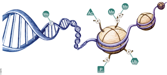
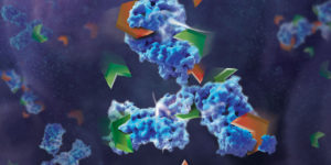
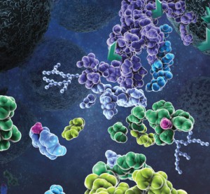
Protein phosphorylation is the most widespread type of post-translational modification. It affects every basic cellular process, including metabolism, growth, division, differentiation, motility, organelle trafficking, membrane transport, muscle contraction, immunity, learning and memory (1,2). Protein kinases catalyse the transfer of the phosphate from ATP to specific amino acids in proteins. In eukaryotes, these are usually Ser, Thr and Tyr residues. Due to the development of specific phosphopeptide enrichment techniques and highly sensitive MS instruments, phosphoproteomics has enabled researchers to gain a comprehensive view on the dynamics of protein phosphorylation and phosphorylation based signaling networks.

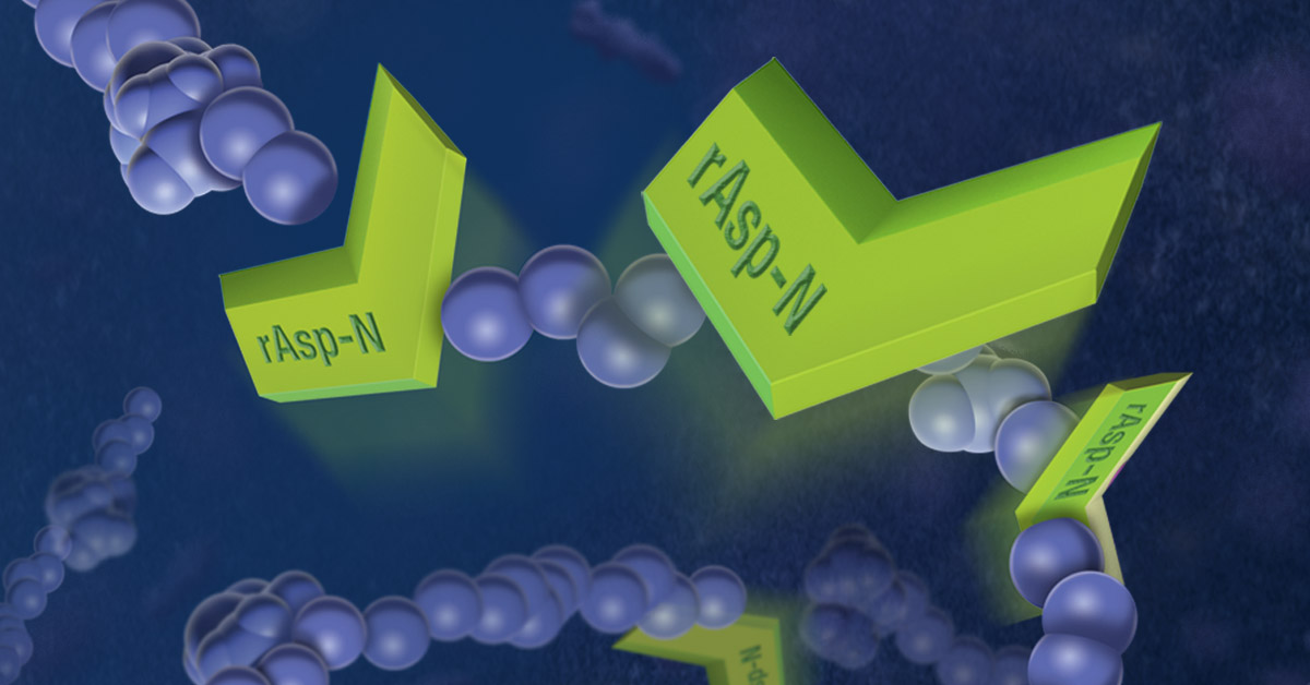
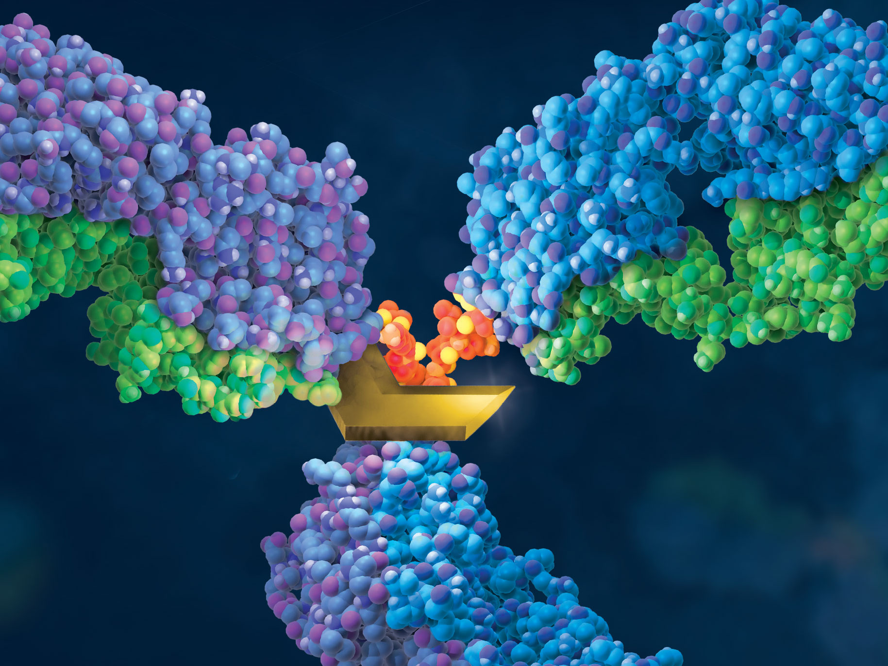
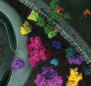
Due to its high cleavage specificity, trypsin is the commonly used proteolytic enzyme in MS-based proteomics, cleaving peptides carboxyterminal of the amino acids lysine and arginine. However, various factors such as the tertiary structure of a protein, adjacent basic amino acids or negatively charged residues close to cleavage sites as well as PTMs are known to impair proteolysis.

To gain closer insights into the impact of phosphorylation on tryptic digestion, a recent publication(3) systematically characterized the digestion efficiency of model peptide sequences that are known to be prone to incomplete digestion.

The results indicated that increasing trypsin concentrations up to a trypsin to peptide ratio of 1:10 led to a significant gain (1) in the overall number of phosphorylation sites (up to 9%) and in the intensities of individual phosphopeptides, thereby improving the sensitivity of phosphopeptide quantification.

The effect of organic solvents (ACN, acetonitrile and TFE trifuorethanol was also evaluated). Positive results were noted with TFE when determining the digestion of individual peptides. However TFE interfered with TiO2 phosphopeptide enrichment and therefore was not recommended for use with complex samples.

  1. Engholm-Keller, K and Larsen, M.R. (2013) Technologies and challenges in large scale phosphoroproteomics. Proteomics 13, 910–31.
  2. Beausoleil, S. A. et al. (2010) Tissue-specific atlas of mouse protein phosphorylation and expression. Cell 143, 1174–89.
  3. Dickhut, C. et al. (2014) Impact of Digestion Conditions on phosphoproteomics. J. Proteome Res. 13, 2761–70.

Use of Mass Spectrometry to Quantitate Food Allergens


Food allergies are becoming increasingly prevalent among children. Credit: James Gathany, CDC

Food allergies are increasing worldwide and becoming a public health issue, especially among children are concerned. Children have a higher prevalence of food allergies, with about 4–8%, compared to adults (1–5%).  Currently antibody-based methods such ELISA (enzyme-linked immunosorbent assay) are the primary method for food allergen analysis. In most cases antibodies are only available for single well-known allergens. Often those that are commercially available are poorly characterized resulting cross-reactivity that leads to false-positive results in diagnostic tests.

A recent publication (1) presented a review of an alternative technology based on mass spec (i.e., multiple reaction monitoring, MRM) that circumvents the drawbacks of antibody based methods. MRM allows precise quantitative determination of target proteins in complex samples with broad dynamic range.  MRM also provides quantification of different isoforms. It is noted that tryptic digestion followed by mass spec analysis, has already identified several unique peptides for different allergens, including those found in crustaceans, eggs, fish, peanuts, soy and wheat. In summary the challenge is now to select the appropriate tryptic signature peptide(s) for the respective allergen and to develop well characterized standards (i.e., isotope labeled standards) to ensure accurate quanititation.

Citation:

Koeberl, M et al. (2014) Next Generation of Food Allergen Quantification using Mass Spectrometric Systems J. Proteome Research  13, 3499–509.