Mass Spec for Glycosylation Analysis of SARS-CoV-2 Proteins Implicated in Host-Cell Entry

The spike protein of the SARS-CoV-2 virus is a very commonly researched target in COVID-19 vaccine and therapeutic studies because it is an integral part of host cell entry through interactions between the S1 subunit of the spike protein with the ACE2 protein on the target cell surface. Viral proteins important in host cell entry are typically highly glycosylated. Looking at the sequence of the SARS-CoV-2 virus, researchers predict that the spike protein is highly glycosylated. In a recent study, researchers conducted a glycosylation analysis of SARS-CoV-2 proteins using mass spec analysis to determine the N-glycosylation profile of the subunits that make up the spike protein.
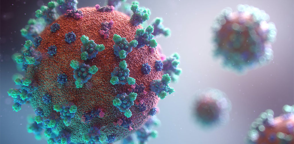
3d model of coronavirus covid-19 showing the spike protein. A recent study performed a glycosylation analysis of SARS-CoV-2 protein.

Glycans assist in protein folding and help the virus avoid immune recognition by the host. Glycosylation can also have an impact on the antigenicity of the virus, as well as potential effects on vaccine safety and efficacy. Mass spectrometry is widely used for viral characterization studies of influenza viruses. Specifically, mass spec has been used to study influenza protein glycosylation, antigen quantification, and determination of vaccine potency.

Continue reading “Mass Spec for Glycosylation Analysis of SARS-CoV-2 Proteins Implicated in Host-Cell Entry”
Understanding the Structure of SARS-CoV-2 Spike Protein

Glycosylation is the process by which a carbohydrate is covalently attached to target macromolecules, typically proteins. This modification serves various functions including guiding protein folding (1,2), promoting protein stability (2), and participating signaling functions (3).

ribbon structure of SARS-CoV-2 protein
Ribbon Structure of SARS-CoV-2 Spike Protein

SARS-CoV-2 utilizes an extensively glycosylated spike (S) protein that protrudes from the viral surface to bind to angiotensin-converting enzyme 2 (ACE2) to mediate host-cell entry. Vaccine development has been focused on this protein, which is the focus of the humoral immune response. Understanding the glycan structure of the SARS-CoV-2 virus spike (S) protein will be critical in the development of glycoprotine-based vaccine candidates.

Continue reading “Understanding the Structure of SARS-CoV-2 Spike Protein”

Bottom-up Proteomics: Need Help?

The use of mass spectrometry for the characterization of individual or complex protein samples continues to be one of the fastest growing fields in the life science market.

Bottom-up proteomics is the traditional approach to address these questions. Optimization of each the individual steps (e.g. sample prep, digestion and instrument performance) is critical to the overall success of the entire experiment.

To address issues that may arise in your experimental design, Promega has developed unique tools and complementary webinars to help you along the way.

Here you can find a summary of individual webinars for the following topics:

Continue reading “Bottom-up Proteomics: Need Help?”

IdeZ Protease: A New Tool for the Characterization of Antibodies

12335MB

Therapeutic monoclonal antibodies are large, complex molecules that undergo numerous post translational modifications (PTMs).  In-depth characterization of antibody PTMs remains a significant hurdle because their large size (~150 kDa) makes mass spectrometry analysis extremely challenging.

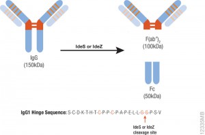
IdeS protease specifically cleaves IgGs into Fab and Fc fragments. This enzyme is highly specific and cleaves human IgG specifically at one site in the lower hinge region.  Because of the exquisite specificity of the enzyme, it produces highly homogeneous Fc and Fab fragments which are then readily analyzed using techniques such as mass spectrometry or HPLC.

Are you looking for proteases to use in your research?
Explore our portfolio of proteases today.

One of the drawbacks of IdeS is that it exhibits poor activity against mouse IgGs. IdeZ Protease is an immunoglobulin-degrading enzyme from Streptococcus equi subspecies zooepidemicus. It is an engineered recombinant protease overexpressed in E. coli. Like IdeS Protease, IdeZ Protease specifically cleaves IgG molecules below the hinge region to yield F(ab′)2 and Fc fragments.  Reduction of the digestion products produces three fragments of ~25kDa that are readily analyzed by LC-MS.

One of the key advantages of the IdeZ Protease is that it has significantly improved activity against mouse IgG2a and IgG3 subclasses compared to IdeS Protease. IdeZ Protease does not cleave mouse IgG1 or IgG2b.

Key technical parameters when digesting mouse IgGs utilizing IdeZ are the following:

• Add 1 unit of IdeZ Protease per 1µg of IgG to be digested.
• IdeZ Protease is most active in buffers at or near neutral pH. The recommended digestion buffer is 50mM sodium phosphate, 150mM NaCl (pH 6.6).
• Mouse IgG2a and IgG3 typically require 2–4 hours at 37°C  for complete digestion.
• IdeZ Protease has a histidine tag for easy removal if so desired.

Monitoring Mass Spec Instrument Performance and Sample Preparation

Proteomics, the analysis of the entire protein content of a living system, has become a vital part of life science research, and mass spectrometry (MS) is the method for analyzing proteins.  MS analysis of protein content allows researchers to identify proteins, sequence them and determine the nature of post translational modifications.

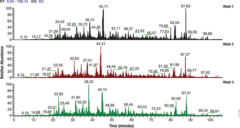
LC/MS performance monitoring. Each run used 1μg of human predigested protein extract injected into the instrument (Waters NanoAquity HPLC System interfaced to a Thermo Fisher Q Exactive™ Hybrid Quadrupole-Orbitrap Mass Spectrometer). Peptides were resolved with a 2-hour gradient. Weekly monitoring with the human extract ensured consistent analytical performance of the instrument.

Mass spectrometry allows characterization of molecules by converting them to ions so that they can be manipulated in electrical and magnetic fields. Basically a small sample (analyte) is ionized, usually to cations by loss of an electron. After ionization, the charged particles (ions) are separated by mass and charge;  the separated particles are measured and data displayed as a mass spectrum. The mass spectrum is typically presented as a bar graph where each peak represents a single charged particle having a specific mass-to-charge (m/z) ratio. The height of the peak represents the relative abundance of the particle. The number and relative abundance of the ions reveal how different parts of the molecule relate to each other.

For the study of large, organic macromolecules, matrix associated laser desorption/ionization (MALDI) or tandem mass spec/collision induced dissociation (MS/MS) techniques are often used to generate the charged particles from the analyte. MS analysis brings sensitivity and specificity to proteome analysis. The technique has excellent resolution and is able to distinguish one ion from another, even when their m/z ratios are similar. Macromolecules are present in extremely different concentrations in the cells, and MS analysis can detect biomolecules across five logs of concentration.

Continue reading “Monitoring Mass Spec Instrument Performance and Sample Preparation”