Protein:DNA Interactions—High-Throughput Analysis

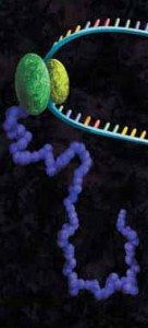
Protein-DNA interactions are fundamental processes in gene regulation in a living cells. These interactions affect a wide variety of cellular processes including DNA replication, repair, and recombination. In vivo methods such as chromatin immunoprecipitation (1) and in vitro electrophoretic mobility shift assays (2) have been used for several years in the characterization of protein-DNA interactions. However, these methods lack the throughput required for answering genome-wide questions and do not measure absolute binding affinities. To address these issues a recent publication (3) presented a high-throughput micro fluidic platform for Quantitative Protein Interaction with DNA (QPID). QPID is an microfluidic-based assay that cam perform up to 4096 parallel measurements on a single device.

The basic elements of each experiment includes oligonucleotides that were synthesized and hybridized to a Cy5-labeled primer and extended using Klenow. All transcription factors that were evaluated contained a 3’HIS and 5’ cMyc tag and were expressed in rabbit reticulocyte coupled transcription and translation reaction (TNT® Coupled Reticulocyte Lysate). Expressed proteins are loaded onto to the QIPD device and immobilized. In the DNA binding assay the fluorescent DNA oligonucleotides are incubated with the immobilized transcription factors and fluorescent images taken. To validate this concept the binding of four different transcription factor complexes to 32 oligonucleotides at 32 different concentrations was characterized in a single experiment. In a second application, the binding of ATF1 and ATF3 to 128 different DNA sequences at different concentrations were analyzed on a single device.

Literature Cited

  1. Ren, B. et al. (2007) Genome-wide mapping of in vivo protein-DNA binding proteins. Science 316, 1497–502.
  2. Garner, M.M. (1981) A gel electrophoresis method for quantifying the binding of proteins to specific DNA regions. Nuc. Acids. Res. 9, 3047-60.
  3. Glick,Y et al. (2016) Integrated microfluidic approach for quantitative high throughput measurements of transcription factor binding affinities. Nuc. Acid Res. 44, e51.
Lost in Translation? Tips for Preparing RNA for in vitro Translation Experiments

In vitro translation of proteins through cell-free expression systems using rabbit reticulocytes, E. coli S30, or wheat germ extracts can be invaluable in studying protein function.  If you only need a small amount (100s of nanograms), it’s also faster and easier than synthesizing vast quantities in bacterial or mammalian cells (~ 90 minutes for cell-free vs. long growth times and extraction steps after an initial optimization for protein synthesized in larger scale).  There are many systems out there, and knowing which to use can sometimes be difficult.  Many kits include components that combine transcription and translation in one-step, eliminating the need to provide your own RNA.  But when you want to make your own RNA templates to add to lysates, then there are additional concerns.

artists concept of in vitro translation
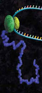
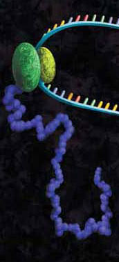
A protein chain being produced from a ribosome.

Many people don’t want to work with RNA since the common lab lore suggests it’s a finicky molecule, and for good reason.  Extracting it requires the utmost care in technique and elimination of nucleases.  Failing to do so results in degradation of the molecule, and so with it your experiments (see our recent blog by Terri Sundquist on tips for isolating RNA with ease).  Preparing RNA for cell-free expression is subject to the same concerns as extracted RNA, but with the proper care is not that much more of a challenge than using a DNA template.

The first step for using cell-free expression systems with RNA templates is to make the RNA.  Here are some tips that will ensure success.

Continue reading “Lost in Translation? Tips for Preparing RNA for in vitro Translation Experiments”

Characterization of Ubiquitination Using Cell-Free Expression

Ubiquitination refers to the post translational modification of a protein by attachment of one or more ubiquitin monomers. The most prominent function of ubiqutin is labeling proteins for proteasome degradation. In addition to this function ubiquitination also controls the stability, function and intracellular localization of a wide variety of proteins.

Cell free expression can be used to characterize ubiquitation of proteins. Target proteins are expressed in a rabbit reticulocyte cell free system (supplemented with E1 ubiquitin activating enzyme, E2 ubiquitin –conjugating enzyme, and ubiquitin). Proteins that have been modified can be analyzed by a shift in migration on polyacrylamide gels.

The following references illustrate the use of cell free expression for this application.

Jung, Y.S. et al. (2011) The p73 Tumor Suppressor Is Targeted by Pirh2 RING Finger E3 Ubiquitin Ligase for the Proteasome-dependent Degradation. J. Biol. Chem. 286, 35388–95.

Su, C-H, et al. (2010) 14-3-3sigma exerts tumor-suppressor activity mediated by regulation of COP1 stability. Cancer. Res. 71, 884–94.

Naoe, H. et al. (2010). The anaphase-promoting complex/cyclosome activator Cdh1 modulates Rho GTPase by targeting p190 RhoGAP for degradation. Mol. Cell. Biol. 30, 3994-05.

de Thonel, A. et al. (2010) HSP27 controls GATA-1 protein level during erythroid cell differentiation. Blood 116, 85–96.

Kaneko, M. et al. (2010) Loss of HRD1-mediated protein degradation causes amyloid precursor protein accumulation and amyloid-beta generation. J. Neurosci. 30, 3924–32.

Cell-Free Kinase Assays

Protein phosphorylation is one of the most biologically relevant modifications and is involved in many eukaryotic and prokaryotic cellular signaling processes. It is estimated that one-third of human proteins are phosphorylated.

The following examples utilize the ability of cell free experession to express active proteins, and when supplemented with the necessary components (e.g., ATP, NaCl), to be used for the characterization of phosphorylation events.

Modrof, J. et al. (2005) Phosphorylation of bluetongue virus nonstructural protein 2 is essential for formation of viral inclusion bodies. J. Vir. 79, 10023–31. Use of TNT® cell-free to express NS2 and NS2 mutant proteins for use in vitro kinase assays to confirm phosphorylation by protein kinase CK2.

Kwon, S. et al. (2005) Signal pathway of hypoxia-inducible factor-1alpha phosphorylation and its interaction with von Hippel-Lindau tumor suppressor protein during ischemia in MiaPaCa-2 pancreatic cancer cells. Clin. Cancer Res. 11, 7607–13. The TNT® system was used to identify which p38 mitogen-activated protein kinase isoform(s) was cabable of phosphorylation of HIF—1 alpha

Harris, J. et al. (2006). Nuclear accumulation of cRel following C-terminal phosphorylation by TBK1/IKK epsilon. J. Immunol. 177, 2527–35. IKK and IKK mutants were expressed using TNT and used in a vitro kinase assay to characterize the recognition motif in cRel transcription domain

Jailais, Y. et al. (2011) Tyrosine phosphorylation controls brassinosteroid receptor activation by triggering membrane release of its kinase inhibitor. Genes Dev. 25, 232–37. Using a vitro kinase assay, full–length and truncations versions of the Brassinostediod-insentive receptor protein were expressed using the TNT® system and incubated with purified BR11 kinase domain to determine binding sites of the two proteins.

Optimized Protein Expression: Flexi Rabbit Reticulocyte Lysate

A protein chain being produced from a ribosome.

mRNAs commonly exhibit differing salt requirements for optimal translation. Small variations in salt concentration can lead to dramatic differences in translation efficiency. The Flexi® Rabbit Reticulocyte Lysate System allows translation reactions to be optimized for a wide range of parameters, including
Mg2+ and K+ concentrations and the choice of adding DTT. To help optimize Mg2+ for a specific message, the endogenous Mg2+ concentration of each lysate batch is stated in the product information included with this product.

The following references utilize the features of Flexi Rabbit Reticulocyte Lysate System to investigate certain parameters of translation:

Vallejos, M. et al. (2010)The 5′-untranslated region of the mouse mammary tumor virus mRNA exhibits cap-independent translation initiation. Nucl Acids Res. 38, 618–32. Identification of internal ribosomal ribosomal entry site in the 5’ untranslated region of the mouse mammary tumor virus mRNA.

Spriggs, K. et al. (2009) The human insulin receptor mRNA contains a functional internal ribosome entry segment. Nucl. Acids. Res. 17, 5881–93. Identification of a functional internal ribosome entry site in the human insulin receptor mRNA.

Powell, M. et al. (2008) Characterization of the termination-reinitiation strategy employed in the expression of influenza B virus BM2 protein. RNA 14, 2394–06. Analysis of the mRNA signals involved in the expression of influenza B virus BM2 protein.

Sato, V. et al. (2007) Measles virus N protein inhibits host translation by binding to eIF3-p40. J. Vir. 81, 11569–76. Charaterized the effect of the measles virus N protein binding to the translation initiation factor eIF3-p40 on the expression of various proteins in rabbit reticulocyte lysate.

Hirao, K. et al. (2006) EDEM3, a soluble EDEM homolog, enhances glycoprotein endoplasmic reticulum-associated degradation and mannose trimming. J. Biol. Chem. 281, 9650–58. The EDEM3 protein was expressed in the presence of canine microsomal membranes to establish that co-translational translocation occurs into the endoplasmic reticulum.

Shenvi, C. et al. (2005) Accessibility of 18S rRNA in human 40S subunits and 80S ribosomes at physiological magnesium ion concentrations–implications for the study of ribosome dynamics. RNA 11, 1898–08. Characterization of ribosome dynamics under different ionic conditions.

Nair, A. et al. (2005) Regulation of luteinizing hormone receptor expression: evidence of translational suppression in vitro by a hormonally regulated mRNA-binding protein and its endogenous association with luteinizing hormone receptor mRNA in the ovary. J. Biol. Chem. 280, 42809–16. Examined the affect of luteinizing hormone receptor mRNA binding protein on transltional suppression of luteinizing hormone receptor RNA.

Cell-Free Applications: RNA Toeprinting

A protein chain being produced from a ribosome.
Precise mapping of the positions of ribosomes and associated factors on mRNAs is essential for characterizing the mechanism of translation. Using the toeprinting assay, mRNA is translated using purified components or crude cell lysates such as rabbit reticulocyte. Cycloheximide is added to the reaction to inhibit elongation. This arrests the position of the ribosomes on the mRNA transcript. The mRNA/ribosomal complex are then copied into cDNA by reverse transcriptase using a complementary radiolabeled primer. Where the reverse transcriptase meets the ribosome bound to the mRNA, cDNA extension is halted, and a toeprint cDNA fragment is generated.

The following references use rabbit reticulocyte lysates as the basis for toeprinting experiments to better understand the mechanism of translation.

Weill, L. et al. (2010)Nucl. Acid, Res. 38, 1367–81. A combination of chemical/enzymatic analyses indicated that gag open reading frame of three viruses adopts a stable secondary structure that allows IRES mediated translation. Mutations that destabilized conserved elements severely inhibit translation. Additional analysis via toeprinting showed HIV-2 IRES has the unique ability to attract up to three initiation complexes on a single RNA molecule.

De Breyne, S. et al. (2008) RNA 14, 367–80. The Simian picornavirus type 9 (SPV9) genome contains a group of IRES that resembles hepacivirus/pestvirus (HP) IRES. Characterization of the initiation process using the toeprinting assay in correlation with other techniques revealed aspects that resemble initiation on the HP IRES and others that are unique to SPV9.

Andreev, D. et al. (2008) RNA 14, 233–39. Rel E is a well characterized toxin involved in the nutritional stress response in bacteria and archae. Rel lacks any eukaryote homolog. Based on toeprinting data, it was demonstrated that RelE cleaves mRNA in the A site of the eukaryote ribosome.

Protease K Protection Assay: Cell Free Expression Application

Microsomal vesicles are used to study cotranslational and initial posttranslational processing of proteins. Processing events such as signal peptide cleavage, membrane insertion, translocation and core glycosylation can be examined by the transcription/translation of the appropriate DNA in the TNT® Lysate Systems when used with microsomal membranes.

The most general assay for translocation makes use of the protection afforded the translocated domain by the lipid bilayer of the microsomal membrane. In this assay protein domains are judged to be translocated if they are observed to be protected from exogenously added protease. To confirm that protection is due to the lipid bilayer addition of 0.1% non-ionic detergent (such as Triton® X-100) solubilizes the membrane and restores susceptibility to the protease.

Are you looking for proteases to use in your research?
Explore our portfolio of proteases today.

Many proteases have proven useful for monitoring translocation in this fashion including Protease K or Trypsin.

The following are examples illustrating this application:

  1. Minn, I. et al. (2009) SUN-1 and ZYG-12, mediators of centrosome-nucleus attachment, are a functional SUN/KASH pair in Caenorhabditis elegans. Mol. Biol. Cell. 20, 4586–95.
  2. Padhan, K. et al. (2007) Severe acute respiratory syndrome coronavirus Orf3a protein interacts with caveolin. J.Gen.Virol. 88, 3067–77.
  3. Tews, B.A. et al. (2007) The pestivirus glycoprotein Erns is anchored in plane in the membrane via an amphipathic helix. J.Biol.Chem. 282, 32730–41.
  4. Pidasheva, S. et al. (2005) Impaired cotranslational processing of the calcium-sensing receptor due to signal peptide missense mutations in familial hypocalciuric hypercalcemia. Hum. Mol. Gen. 14, 1679–90.
  5. Smith, D. et al. (2002) Exogenous peptides delivered by ricin require processing by signal peptidase for transporter associated with antigen processing-independent MHC class I-restricted presentation. J. Immun. 169, 99–107.

Optimized Wheat Germ Extract for High-Yield Protein Expression of Functional, Soluble Protein

Wheat Germ Extract for high-yield protein expression

Cell-free protein synthesis has emerged as powerful alternative to cell based protein expression for functional and structural proteomics. The TNT® SP6 High-Yield Protein Expression System uses a high-yield wheat germ extract supplemented with SP6 RNA polymerase and other components. Coupling transcriptionaland translational activities eliminates the inconvenience of separate in vitro transcription and purification steps for the mRNA, while maintaining the high levels of protein expression (1).

Continue reading “Optimized Wheat Germ Extract for High-Yield Protein Expression of Functional, Soluble Protein”

Cell-Free Protein Synthesis

Cell-free protein synthesis (aka: in vitro translation) refers to protein production in vitro using lysates that provide the cellular machinery necessary for synthesis. Ribosomes, tRNAs, aminoacyl-tRNA synthetases, initiation/elongation/termination factors, GTP, ATP, Mg2+ and K+ are among the requirements for a translation system. These are provided by lysates, which can be from prokaryotic or eukaryotic sources, depending on your requirements.

Cell-free protein synthesis is most commonly used for generating protein for study of things like:

Continue reading “Cell-Free Protein Synthesis”