Avoid False Hits During Compound Screening for Drug Discovery

One goal of drug discovery and research programs is to reduce false hits as early as possible in the process. Follow-up on false hits is costly in terms of time and resources, and the longer the false hits remain in the drug development pipeline, the more costly they are. So methods that can easily reduce the number of false hits during compound screening early in the discovery process are particularly sought after.

Reporter assays have proven to be invaluable tools for elucidating the mechanisms of action of small molecules or other agents on signaling pathways within cells, and the luciferase reporter assay has become a standard research tool in the biological research laboratory.

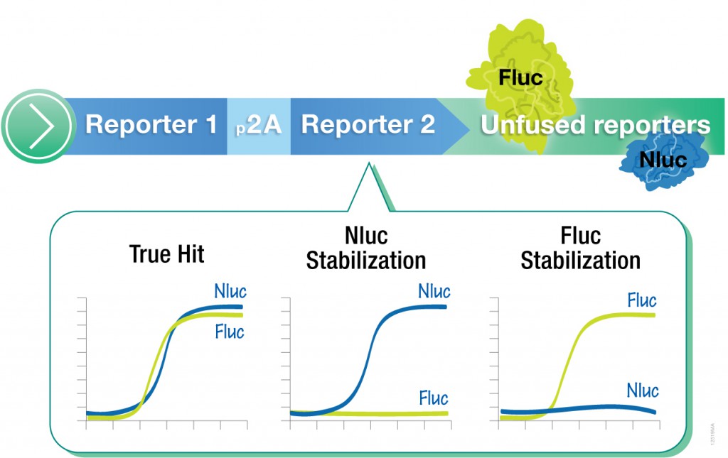
However, one caveat of using standard luciferase-based reporter assays for larger-scale compound screening efforts is the frequency of false hits that result from direct interaction of compounds with the luciferase reporter. This issue can be mitigated with a “coincidence reporter” system where two independent reporter proteins are produced from a single transcript. In this type of assay, a bicistronic transcript is stoichiometrically translated into two nonhomologous reporters by means of a 2A “ribosomal skipping” sequence. Since it is unlikely that compounds will interact with two distinct types of reporter, “coincident” responses will indicate on-target activity. Such a coincident reporter system provides an important control against costly false hits early in drug discovery research programs.

A paper published online in ACS Chem Biol in February describes the first successful application of the firefly/NanoLuc luciferase coincidence reporter system to identify new pathways that up-regulate PARK2 expression.

Continue reading “Avoid False Hits During Compound Screening for Drug Discovery”

Improving Cancer Drug Screening with 3D Cell Culture

Differential contrast image of HCT116 colon cancer spheroid grown in a 96-well hanging-drop platform after seeding with 800 cells. Copyright Promega Corporation.
Tissue culture using primary or cultured cell lines has long been a mainstay of testing compounds for inhibiting cell growth or promoting apoptosis during screening for cancer drugs. However, the standard culture conditions result in monolayers of cells, dividing and growing across the bottom of a well, plate or flask in a single layer. The drawback of this technique is that organisms do not come in monolayers; a three-dimensional (3D) spheroid is closer to the in vivo state, especially if the spheroids are made up of more than one cell type like tumors in multicellular organisms. Even more beneficial would be using 3D cultured cells in high-throughput screening to facilitate compound profiling for target effectiveness and cytotoxicity. In a recent PLOS ONE article, researchers used normal and breast cancer cells both in monoculture and coculture to test a set of compounds and found results differed between 2D and 3D cultured cells. Continue reading “Improving Cancer Drug Screening with 3D Cell Culture”

Compound Screening Using Cell-Free Protein Expression Systems

A protein chain being produced from a ribosome.
Both prokaryotic and eukaryotic cell-free protein expression systems have found great utility in efforts to screen organic compounds for inhibition of the basic cellular functions of transcription and translation, common targets for antibiotic compounds.

Cell-free systems can provide some advantages over cell-based systems for screening purposes. Cell-free systems allow exact manipulation of compound concentrations. This is an important parameter when evaluating the potential potency of the lead compound.

There is no need for cellular uptake to evaluate the effect of the compounds. While uptake evaluation is important for determining the eventual efficacy of the drug, it can unnecessarily eliminate valuable lead compounds in an initial screen. The interpretation of results in living cells is complicated by the large number of intertwined biochemical pathways and the ever-changing landscape of the growing cell. Cell-free systems allow the dissection of effects in a static system for simpler interpretation of results and the ability to specifically monitor individual processes such as transcription or translation. Individual targets not normally present, or found at low concentrations, can be added in controlled amounts.

The following references illustrate this application: