Out-FOXOing High-Stage Neuroblastoma

Fluorescence microscopy of neuroblastoma cells.

In recent years, scientists have been hot on the trail of transcription factor FOXO3, tracing its involvement in various tumor-centric activities comprising the many trademarks of cancer, from drug resistance to metastasis to tumor angiogenesis.

FOXO3 is a member of the O sub-class of the forkhead box family of transcription factors. The forkhead box (FOX) family is characterized by a fork head DNA-binding domain (DBD), comprised of around 100 amino acids. They have also proven themselves to be a family of many hats, functioning in diverse roles ranging from metabolism, immunology, cell-cycle control, development, as well as cancer (1). The forkhead box O (FOXO) sub-class alone has demonstrated involvement in a variety of cellular outcomes, from drug resistance and longevity to apoptosis induction.

Due to its pro-apoptotic and anti-proliferative proclivity, FOXO3 has been previously identified as a tumor suppressor gene. However, more and more studies have begun to flip the narrative on FOXO3, portraying it more as a devoted henchman, due to its roles in drug and radiotherapy resistance, cell-cycle arrest and long-term maintenance of leukemia-initiating stem cells in a variety of cancer types, including breast cancer, pancreatic cancer, glioblastoma, and both acute and chronic myeloid leukemia.

Continue reading “Out-FOXOing High-Stage Neuroblastoma”

NLRP3: The New Hope for Treating Chronic Inflammatory Diseases

Inflammasome - inflammatory diseases caused by NLRP3

Our innate immune system was meant to do good. Up until a century ago, most humans died from infectious diseases like diarrhea, tuberculosis and meningitis. Over millions of years, our immune system has evolved to fight these life-threatening infections from pathogens. As a result, we have developed a highly efficient response to these tiny invaders. But it seems that our immune system may be turning against us.

Continue reading “NLRP3: The New Hope for Treating Chronic Inflammatory Diseases”

Bacteria and Viruses as Cancer Treatments

Over a hundred years ago William B Coley, the “Father of Immunotherapy”, discovered that injection of bacteria or bacterial toxins into tumors could cause those tumors to shrink. The introduction of bacteria had the side-effect of stimulating the immune system to attack the tumor. The field of cancer immunotherapy research—which today includes many different approaches for generating anti-tumor immune responses—originated with these early experiments.

Use of bacteria is one way to stimulate the immune system to attack cancer cells, others include use of cytokines, immune checkpoint blockades and vaccines. This Nature animation provides a simple overview of these methods.

Continue reading “Bacteria and Viruses as Cancer Treatments”

Because Timing Is Everything….

Camp Indian Trails Morning Flag Ceremony
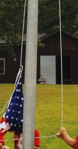
Timing is everything! I learned that the hard way just two weeks ago when I took my son to scout camp and thought I would try to capture the traditional American flag ceremony for posterity. I set up my camera for a panoramic shot and scanned the crowd.  Feeling very pleased with myself, I got home that evening ready to show my family the great camera skills I had honed over the Summer months. To my horror, I noticed that half of the scout troop was saluting the flag while the other half were standing to attention! I had got the timing horribly wrong (although the picture is still fun to look at in a strange sort of way).

Timing is everything in science as well.  As a technical services scientist at Promega I have sung the ‘timing’ tune to many a biologist. No more so than in the study of apoptosis where Caspases activate each other in a choreographed cascade of molecular triggers that all have their place and time in a domino sequence of enzymatic cleavage events. I frequently talk to researchers about that ‘sweet spot’ of activity when any given Caspase is busily cleaving a peptide moiety off of the next Caspase in the sequence. Finding that sweet spot is anything but trivial and often requires a considerable amount of patience during the optimization phase of experimentation.

Promega has developed a comprehensive suite of systems (see here) designed to help get the timing right for the cell and compound combinations you might be working with.  The end result is that you have experiments that are timed so as to give you reliable information about what is really happening in your cells.

Detecting Apoptosis

The concept of cell death as a normal cell fate was articulated only three years after Schleiden and Schwann introduced the Cell Theory when, in 1874, Vogt described natural cell death as an integral part of toad development (as cited in Cotter and Curtin, 2003). Since these early observations, natural cell death has been described “anew” several times. In 1885 Flemming provided the first morphological description of a natural cell death process, which we now label “apoptosis”, a term coined by Kerr and colleagues to describe the unique morphology associated with a cell death that differs from necrosis (as cited in Kerr et al. 1972).

Apoptosis morphology
Progression of morphology changes during apoptosis.

In the 1970s and 1980s, studies revealed that apoptosis not only had specific morphological characteristics but that it was also a tightly regulated process with specific biochemical characteristics. Studies of cell lineage in the nematode, Caenorhabditis elegans, showed that apoptosis was a normal feature of the nematode’s invariant developmental program (Hengartner, 1997). At the biochemical level, Wyllie showed that DNA degradation by a specific endonuclease during apoptosis resulted in a DNA ladder composed of mono- and oligonucleosomal-sized fragments (Wyllie, 1980).

These and many other studies have proven that apoptosis is a critical component of development, and when it doesn’t happen appropriately, it can be pathological, leading to cancers or other diseases. Therefore, understanding how and when apoptosis occurs and the many signals that can trigger this process is a focus of many laboratory experiments.

There are many ways to detect apoptosis in cells or tissues. This blog describes some of the most common ones.

Continue reading “Detecting Apoptosis”

Shining a Bright Light on Deep Questions in Biology with Bioluminescence

artists view inside a cell

Search the PubMed database for “dual-luciferase” and citations abound. The Dual-Luciferase® Reporter Assay is a powerful tool that allows researchers to ask a multitude of questions about gene control and expression in a system that itself could be normalized and internally controlled. For more than 15 years, firefly and Renilla luciferases  have formed the basis of a range of powerful assays and research tools for scientists who are asking questions about the deep and complex genetic and cellular story associated with cancer. Here we talk a bit of about bioluminescent chemistries, some of the newest bioluminescent tools available, and how some of these tools can be used to probe the deeper questions of cell biology, including cancer biology.

Continue reading “Shining a Bright Light on Deep Questions in Biology with Bioluminescence”