Image adapted from original artwork by iSO-FORM LLC.
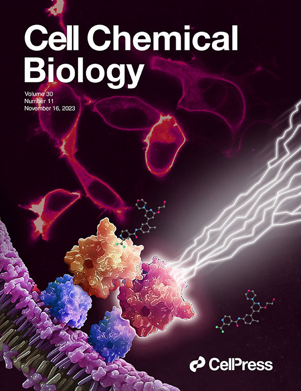
We made the cover! Of Cell Chemical Biology, that is.
This July, Cell Chemical Biology editors accepted a study from Promega scientists and invited the research team to submit cover art for the issue. The study in question details a BRET-based method to quantify drug-target occupancy within RAF-KRAS complexes in live cells. Promega scientists Matt Robers and Jim Vasta collaborated with one of our talented designers, Michael Stormberg, to craft an image that accurately represents the science in a dynamic and engaging way.
I spoke with Michael Stormberg to learn more about the creative process that went into creating this cover art and how he worked with the research team and other collaborators.
Antibody tests are often used to determine whether individuals have been exposed to certain bacteria or viruses. For most existing antibody tests, the process goes something like this: A vial of blood is drawn from the individual, the vial is sent to a lab, then a trained technicians performs the antibody test and sends back the results. The current process is less than ideal for a few reasons. For one, blood draws are invasive and can be painful. Also, getting results could take days, due to the time required to deliver and process the sample. Lastly, costs can be high, since the need for trained professionals and specialized instruments in laboratory settings adds to the cost of each test.
What if all you needed to do for an antibody test was apply a single drop of blood onto a thin piece of film, and you would get results on the spot within five minutes? Scientists have recently developed an antibody test based on bioluminescent technology that could make this a reality. They describe their findings in a recent study published in ACS Sensors.
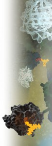
What can you do with a small, super bright luciferase? Amazing things. We’ve highlighted many of the papers and new applications that NanoLuc® luciferase has enabled on this blog. While NanoLuc® luciferase was first introduced as a reporter enzyme to assess promoter activity, its capabilities have expanded far beyond a genetic reporter, creating bioluminescent tools used to study endogeneous protein dynamics, target engagement, protein degradation, immunodetection and more. So where did the NanoLuc luciferase come from and how does one enzyme power so many research capabilities? Read further for a primer on the various technologies and applications developed from this enzyme over the last 10 years.
Researchers having been sharing plasmids ever since there were plasmids to share. Back when I was in the lab, if you read a paper and saw an interesting construct you wished to use, you could either make it yourself or you could “clone by phone”. One of my professors was excellent at phone cloning with labs around the world and had specific strategies and tactics for getting the plasmids he wanted. Addgene makes this so much easier to share your constructs from lab to lab. Promega supports the Addgene mission statement: Accelerate research and discovery by improving access to useful research materials and information. Many of our technology platforms like HaloTag® Fusion Protein, codon-optimized Firefly luciferase genes (e.g., luc2), and NanoLuc® Luciferase are present in the repository. We encourage people to go to Addgene to get new innovative tools. Afterall, isn’t science better when we share?
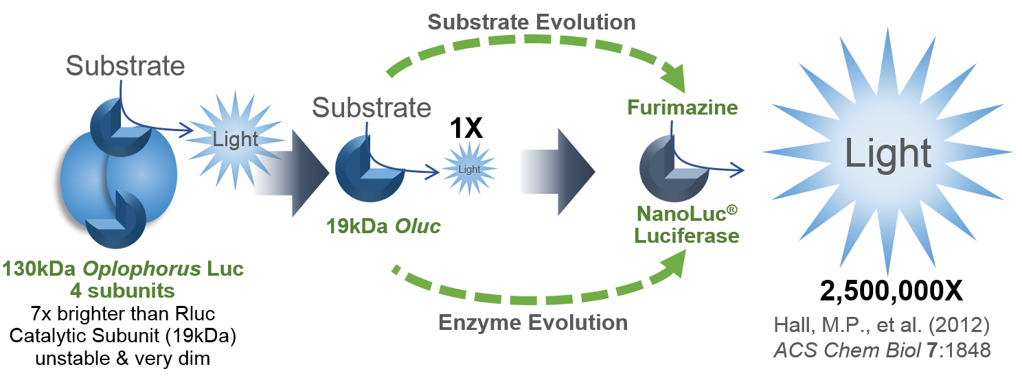
I’d like to focus on some tools in the Addgene collection based on NanoLuc® Luciferase (NLuc). The creation of NanoLuc® Luciferase and the optimal substrate furimazine is a good story (1). From a deep sea shrimp to a compact powerhouse of bioluminescence, NLuc is 100-fold brighter than our more common luciferases like firefly (FLuc) and Renilla (RLuc) luciferase. This is important not so much for how bright you can make a reaction but for how sensitive you can make a reaction. NLuc requires 100-fold less protein to produce the same amount of light from a Fluc or RLuc reaction. NLuc lets you work at physiological concentrations. NLuc is bright enough to detect endogenous tagged genes generated through the CRISPR/Cas9 knock-in. NLuc is very inviting for endogenous tagging as it is only 19kDa. An example is the CRISPaint-NLuc construct (Plasmid #67178) for use in the system outlined in Schmid-Burgk, J.L. et al (2).
Fluorescence resonance energy transfer (FRET) probes or sensors are commonly used to measure cellular events. The probes typically have a matched pair of fluorescent proteins joined by a ligand-binding or responsive protein domain. Changes in the responsive domain are reflected in conformational changes that either bring the two fluorescent proteins together or drive them apart. The sensors are measured by hitting the most blue-shifted fluorescent protein with its excitation wavelength (donor). The resulting emission is transferred to the most red-shifted fluorescent protein in the pair, and the result is ultimately emission from the red-shifted protein (acceptor).
As pointed out by Aper, S.J.A. et al. below, FRET sensors face challenges of photobleaching, autofluorescence, and, in the case of exciting cyan-excitable donors, phototoxicity. Another challenge to using FRET sensors comes when employing optogenetic regulators to initiate the event you wish to monitor. Optogenetic regulators respond to specific wavelengths and initiate signaling. The challenge comes when the FRET donor excitation overlaps with the optogenetic initiation wavelengths. Researchers have sought to alleviate many of these challenges by exchanging the fluorescent donor for a bioluminescent donor, making bioluminescence resonance energy transfer (BRET) probes. In the three papers described below, the authors chose NanoLuc® Luciferase as the BRET donor due to its extremely bright signal.
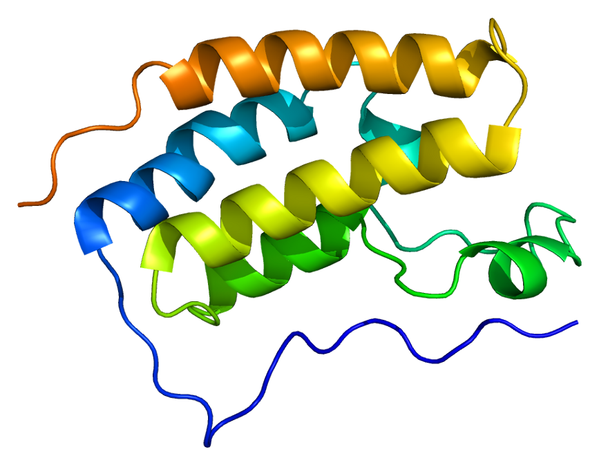
“Protein BRD4 PDB 2oss” by Emw – Own work. Licensed under CC BY-SA 3.0 via Wikimedia Commons – https://commons.wikimedia.org/wiki/File:Protein_BRD4_PDB_2oss.png#/media/File:Protein_BRD4_PDB_2oss.png
One of the more exciting reporter molecules technologies available came online in the past year, with the launch of the Promega NanoBRET™ technology. While it’s easy for me, a science writer at Promega, to brag, seriously, this is a very cool protein interactions tool.
A few of the challenges facing protein-protein interactions researchers include:
The ability to quantitatively characterize protein-protein interactions
Ability to examine protein-protein interactions in situ, in the context of the living cell
A goal of the NanoBRET™ developers was to improve the sensitivity and dynamic range of traditional BRET technology, in order to address these challenges.
In May 2015 these researchers published an article outlining their efforts to create NanoBRET technology in ACS Chemical Biology, in an article entitled, “NanoBRET—A Novel BRET Platform for the Analysis of Protein-Protein Interactions”. Here is a brief look at their work.
Robert Hooke first coined the term “cell” after observing plant cell walls through a light microscope—little empty chambers, fixed in time and space. However, cells are anything but fixed.
Cells are dynamic: continually responding to a shifting context of time, environment, and signals from within and without. Interactions between the macromolecules within cells, including proteins, are ever changing—with complexes forming, breaking up, and reforming in new ways. These interactions provide a temporal and special framework for the work of the cell, controlling gene expression, protein production, growth, cell division and cell death.
Visualizing and measuring protein:protein interactions at the level of the cell without perturbing them is the goal of every cell biologist.
A recent article by Thomas Machleidt et al. published in ACS Chemical Biology, describes a new technology that brings us closer to being able to realize that goal.
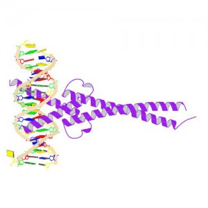
Crystal Structure of MYC MAX Heterodimer bound to DNA ImageSource=RCSB PDB; StructureID=1nkp; DOI=http://dx.doi.org/10.2210/pdb1nkp/pdb;
In 1982, picked up because of its homology to chicken virus genes that could transform cells, MYC became one of the first human genes identified that could drive cellular transformation (1,2). Since that time countless laboratories have prodded and poked the human MYC gene, the MYC protein, their homologs in other animal models, and their transforming viral counterparts.
MYC is a transcription factor and forms heterodimers with a required protein partner, MAX, before binding to the E box sequences of DNA regulatory regions (3). MYC regulates gene expression of many targets through interactions with a host of proteins, often referred to as the MYC Interactome (2). In fact, MYC is estimated to bind 10–15% of the genome, and it regulates the expression of genes that are transcribed by by each of the three RNA polymerases (2).
MYC plays a central role in regulating cell growth, proliferation, apoptosis, differentiation and transformation, acting as a central integrator of cellular signals. MYC is tightly regulated at multiple levels from gene expression to protein stability. Dysregulation (usually upregulation) of the amount and stability of Myc protein is observed in many human cancers. Even in cancers in which MYC is not directly involved in transforming cells, its normal expression is often required to support the extracellular matrix and/or vascularization necessary for tumor growth and formation (4).
Because MYC is such a central player cancer pathology, it is an attractive target for cancer therapeutics (2) .
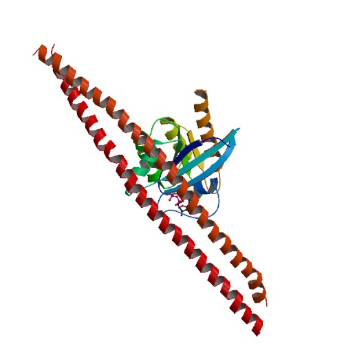
In a study published in Proceedings of the National Academy of Sciences USA article, Wang et al. used the principle of the Promega NanoBRET™ assay to understand how ERK1/2 phosphorylation of Rabin8, a guanine nucleotide exchange factor, influenced its configuration and subsequent activation of Rab8, a protein that regulates exocytosis.
Crystal structure of GDP-boudn Rab8:Rabin8 ImageSource=RCSB PDB; StructureID=4lhy; DOI=http://dx.doi.org/10.2210/pdb4lhy/pdb;
Rab8 is a member of the Rab family of small GTPases and an important regulator of membrane trafficking from the trans Golgi network and recycling endosomes to the plasma membrane. Wang et al. were interested in learning how the guanine nucleotide exchange factor (GEF) Rabin8, a known activator of Rab8, was itself activated to better understand how Rab8 and exocytosis were regulated in the cell. First, they confirmed if the consensus extracellular-signal-regulated kinases ERK1/2 phosphorylation motif uncovered in Rabin8 resulted in phosphorylation of Rabin8. Both in vitro analysis and cell-based assays confirmed that ERK1/2 phosphorylated Rabin8. Next, the GEF activity of Rabin8 was assessed to determine if ERK1/2 phosphorylation activated the GEF. Researchers confirmed activation of Rabin8 GEF in vitro.
XWe use cookies and similar technologies to make our website work, run analytics, improve our website, and show you personalized content and advertising. Some of these cookies are essential for our website to work. For others, we won’t set them unless you accept them. To learn more about our approach to Privacy we invite you to Read More
By clicking “Accept All”, you consent to the use of ALL the cookies. However you may visit Cookie Settings to provide a controlled consent.
We use cookies and similar technologies to make our website work, run analytics, improve our website, and show you personalized content and advertising. Some of these cookies are essential for our website to work. For others, we won’t set them unless you accept them. To find out more about cookies and how to manage cookies, read our Cookie Policy.
If you are located in the EEA, the United Kingdom, or Switzerland, you can change your settings at any time by clicking Manage Cookie Consent in the footer of our website.
Necessary cookies are absolutely essential for the website to function properly. These cookies ensure basic functionalities and security features of the website, anonymously.
Cookie
Duration
Description
cookielawinfo-checbox-analytics
11 months
This cookie is set by GDPR Cookie Consent plugin. The cookie is used to store the user consent for the cookies in the category "Analytics".
cookielawinfo-checbox-functional
11 months
The cookie is set by GDPR cookie consent to record the user consent for the cookies in the category "Functional".
cookielawinfo-checbox-others
11 months
This cookie is set by GDPR Cookie Consent plugin. The cookie is used to store the user consent for the cookies in the category "Other.
cookielawinfo-checkbox-advertisement
1 year
The cookie is set by GDPR cookie consent to record the user consent for the cookies in the category "Advertisement".
cookielawinfo-checkbox-necessary
11 months
This cookie is set by GDPR Cookie Consent plugin. The cookies is used to store the user consent for the cookies in the category "Necessary".
cookielawinfo-checkbox-performance
11 months
This cookie is set by GDPR Cookie Consent plugin. The cookie is used to store the user consent for the cookies in the category "Performance".
gdpr_status
6 months 2 days
This cookie is set by the provider Media.net. This cookie is used to check the status whether the user has accepted the cookie consent box. It also helps in not showing the cookie consent box upon re-entry to the website.
lang
This cookie is used to store the language preferences of a user to serve up content in that stored language the next time user visit the website.
viewed_cookie_policy
11 months
The cookie is set by the GDPR Cookie Consent plugin and is used to store whether or not user has consented to the use of cookies. It does not store any personal data.
Analytical cookies are used to understand how visitors interact with the website. These cookies help provide information on metrics the number of visitors, bounce rate, traffic source, etc.
Cookie
Duration
Description
SC_ANALYTICS_GLOBAL_COOKIE
10 years
This cookie is associated with Sitecore content and personalization. This cookie is used to identify the repeat visit from a single user. Sitecore will send a persistent session cookie to the web client.
vuid
2 years
This domain of this cookie is owned by Vimeo. This cookie is used by vimeo to collect tracking information. It sets a unique ID to embed videos to the website.
WMF-Last-Access
1 month 18 hours 24 minutes
This cookie is used to calculate unique devices accessing the website.
_ga
2 years
This cookie is installed by Google Analytics. The cookie is used to calculate visitor, session, campaign data and keep track of site usage for the site's analytics report. The cookies store information anonymously and assign a randomly generated number to identify unique visitors.
_gid
1 day
This cookie is installed by Google Analytics. The cookie is used to store information of how visitors use a website and helps in creating an analytics report of how the website is doing. The data collected including the number visitors, the source where they have come from, and the pages visted in an anonymous form.
Advertisement cookies are used to provide visitors with relevant ads and marketing campaigns. These cookies track visitors across websites and collect information to provide customized ads.
Cookie
Duration
Description
IDE
1 year 24 days
Used by Google DoubleClick and stores information about how the user uses the website and any other advertisement before visiting the website. This is used to present users with ads that are relevant to them according to the user profile.
test_cookie
15 minutes
This cookie is set by doubleclick.net. The purpose of the cookie is to determine if the user's browser supports cookies.
VISITOR_INFO1_LIVE
5 months 27 days
This cookie is set by Youtube. Used to track the information of the embedded YouTube videos on a website.
Performance cookies are used to understand and analyze the key performance indexes of the website which helps in delivering a better user experience for the visitors.
Cookie
Duration
Description
YSC
session
This cookies is set by Youtube and is used to track the views of embedded videos.
_gat_UA-62336821-1
1 minute
This is a pattern type cookie set by Google Analytics, where the pattern element on the name contains the unique identity number of the account or website it relates to. It appears to be a variation of the _gat cookie which is used to limit the amount of data recorded by Google on high traffic volume websites.