Characterizing Compound Binding in Cell-Free Systems

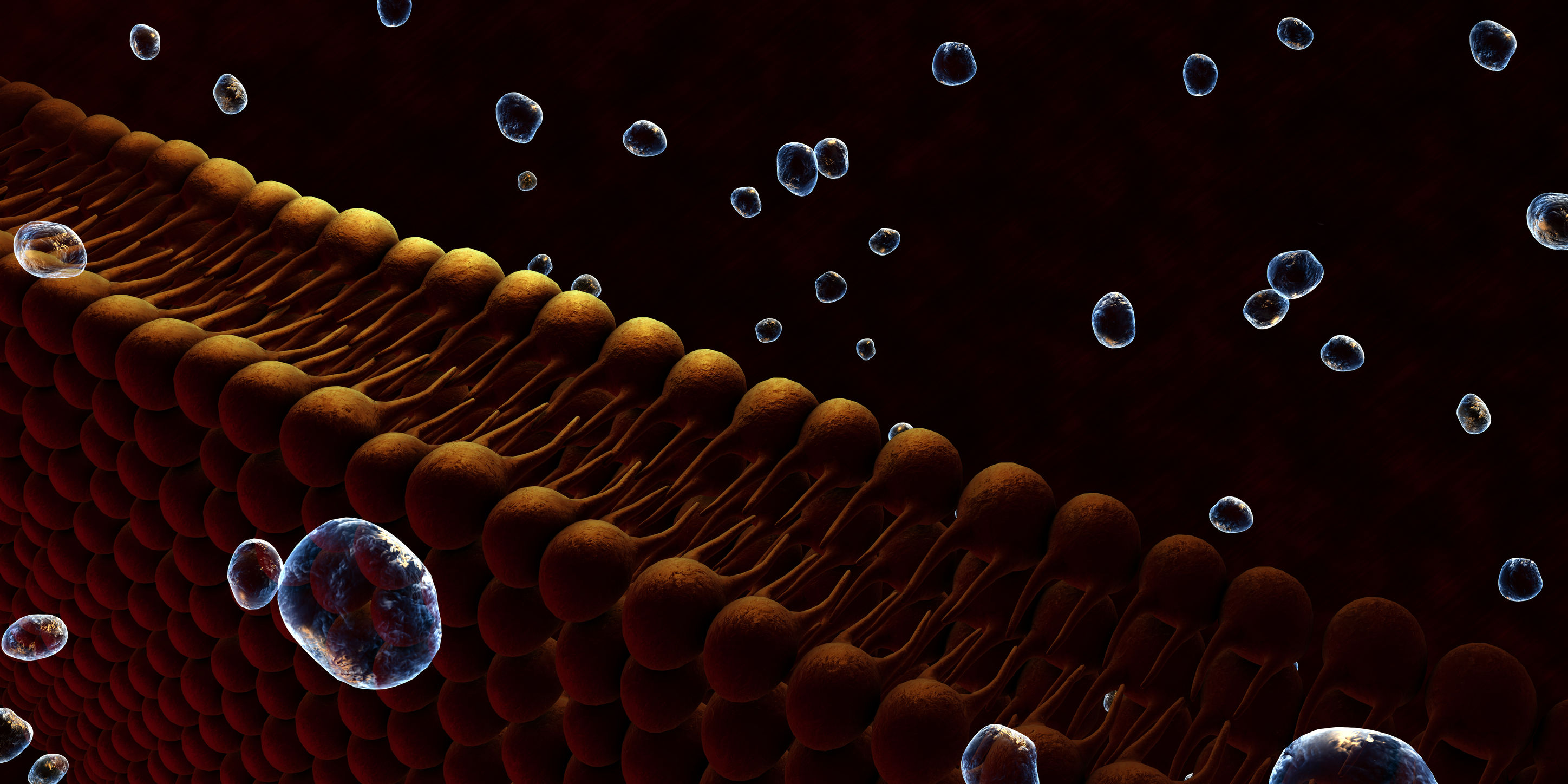
Dioxins (e.g., 2,3,7,8-Tetrachlorodibenzo-p-dioxin, TCDD) and related compounds (DRCs) are persistent environmental pollutants that gradually accumulate through the food chain, mainly in the fatty tissues of animals. Dioxins are highly toxic and can cause reproductive and developmental problems, damage the immune system, interfere with hormones and also cause cancer. This broad range of toxic and biological effects of DRCs is mostly mediated by the aryl hydrocarbon receptor (AHR).

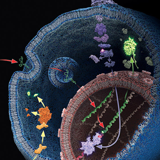
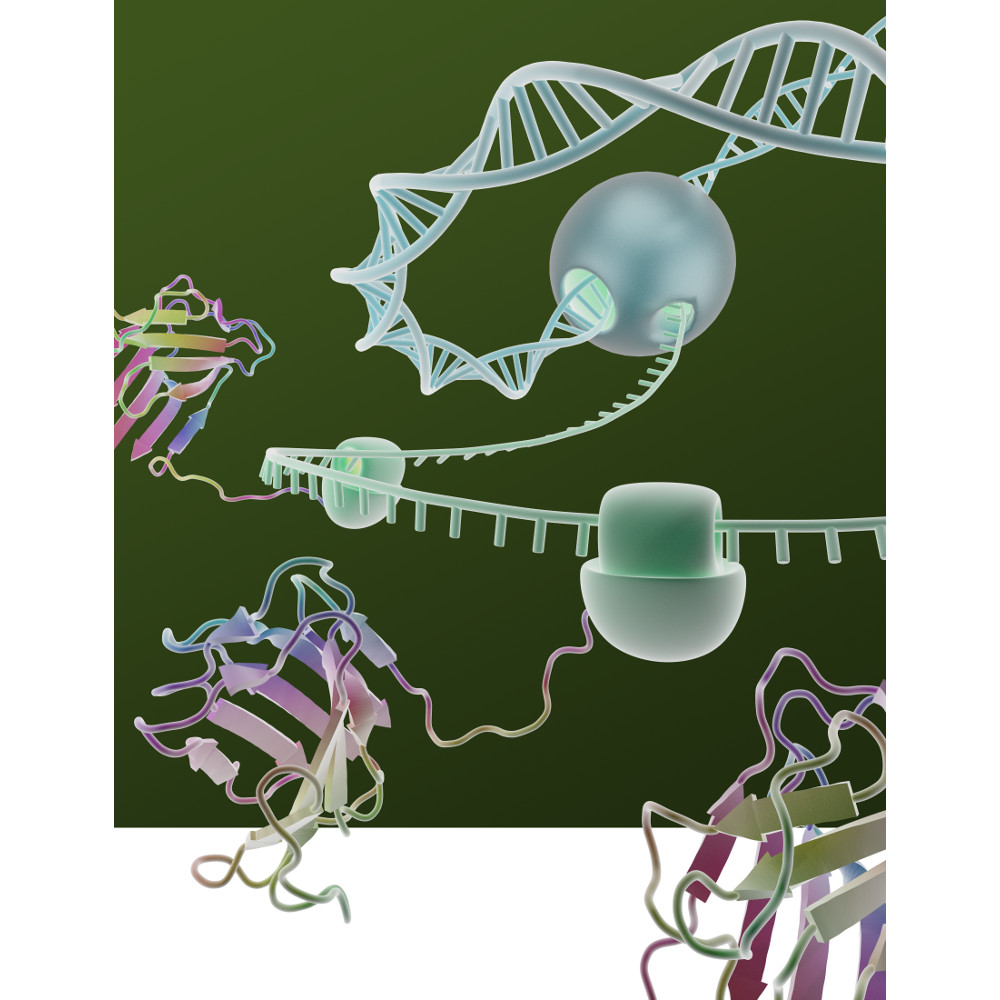
In animal cells, DRCs bind to AHR in the cytoplasm and then translocate into the nucleus, where they affect the transcription of multiple target genes, including xenobiotic-metabolizing enzymes, such as CYP1A isozymes. AHR is also involved in immune system maintenance, protein degradation and cell proliferation.

The jungle crow (Corvus macrorhynchos) has been considered a suitable indicator for monitoring environmental chemicals such as DRCs. While mammals only have one AHR form, avian species have multiple AHR isoforms such as AHR1 and AHR2. To unveil the functional diversity of multiple avian AHR isoforms in terms of their contribution to responses to DRCs a recent study by Kim et al. investigated the molecular and functional characteristics of jungle crow AHR isoforms, cAHR1 and jcAHR2 (1).

cAHR1 and jcAHR2 proteins were synthesized using AHR proteins were synthesized using the TnT Quick-Coupled Reticulocyte Lysate System  to examine whether these jcAHRs have the potential to bind to TCDD. TCDD-binding affinity of the in vitro-expressed jcAHR protein was analyzed using the velocity sedimentation assay with a sucrose gradient.

The results demonstrate that both jcAHR1and jcAHR2 are capable of binding to TCDD.

Reference
Kim, E-Y (2019) The aryl hydrocarbon receptor 2 potentially mediates cytochrome P450 1A induction in the jungle crow (Corvus macrorhynchos). Ecotoxicology and Environmental Safety 171. 99–111

Mutation Analysis Using HaloTag Fusion Proteins

In a recent reference, Kinoshita and colleagues characterized the phosphorylation dynamics of MEK1 in human cells by using the phosphate affinity electrophoresis technique, Phos-tag sodium dodecyl sulfate–polyacrylamide gel electrophoresis (Phos-tag SDS-PAGE; 1). They found that multiple variants of MEK1 with diferent phosphorylation states are constitutively present in typical human cells.

To investigate the relationships between kinase activity and drug efficacy researchers from the same laboratory group conducted phosphorylation profling of various MEK1 mutants by using Phos-tag SDS- PAGE (2).

They introduced mutations in of the MEK-1 coding gene that are associated with spontaneous melanoma, lung cancer, gastric cancer, colon cancer and ovarian cancer were introduced into Flexi HaloTag clone pFN21AE0668, which is suitable for expression of N-terminal HaloTag-fused MEK1 in mammalian cells. Continue reading “Mutation Analysis Using HaloTag Fusion Proteins”

Optimizing Pressure Cycling Sample Preparation for Bottom-Up Proteomics

Large-scale analyses of the proteome have revealed proteomic changes in response to disease, and these changes hold great promise for diagnostics and treatment of complex diseases if proteomic analysis can be brought into the clinical laboratory. Successful and reliable large-scale proteomics requires sample preparation workflows that are reproducible, reliable and show little variability. To bring proteomics into the clinical laboratory, standardized procedures and workflows for sample prep and analysis are required to generate valid, actionable results on a time scale useful for the clinic.

The two most common sample types analyzed for clinical proteomics are body fluids and tissue biopsies. To process these kinds of samples, there are two initial steps: tissue solubilization, followed by proteolytic digestion. Solubilization of solid tissues is the most labor-intensive and produces the most variable results.

The introduction of pressure cycling technology (PCT) using Barocycler instrumentation has greatly improved both tissue solubilization and digestion consistency. The PCT-based sample preparation protocols generally utilize urea as a lysis buffer for protein denaturing and solubilization. Urea has several drawbacks including inhibiting trypsin activity and introducing unwanted modifications like carbamylation.

Lucas and colleagues analyzed whether replacing urea with SDC would produce similar tissue digestion profiles and improve the PCT method.

Are you looking for proteases to use in your research?
Explore our portfolio of proteases today.

SDC allowed the use of higher temperatures compared to urea, and hence the first step (lysis, reduction, and alkylation) was performed at 56 °C. The second digestion step in the Barocycler was optimized, and the third step was eliminated. To further reduce digestion time, they capitalized on Rapid Trypsin/Lys-C.  Rapid Trypsin/Lys-C maintains robust activity at 70 °C, and allowed Barocycler digestion to be performed in a single step, completing digestion in 30 cycles (approximately 30 min) rather than 105 minutes, streamlining the protocol.

The data presented an improved conventional tissue PCT approach in a Barocycler by replacing urea and proteolytic enzymes with SDC, N-propanol, and modified commercially available enzymes that have higher optimum temperatures.


Try a sample of high-efficiency Trypsin Platinum today!

Visit our website for more on Trypsin Platinum, Mass Spectrometry Grade, with enhanced proteolytic efficiency and superior autoproteolytic resistance.


Paper Referenced

Lucas, N. et al. (2019) Accelerated Barocycler Lysis and Extraction Sample Preparation for Clinical Proteomics by Mass Spectrometry. J of Proteome Res 18, 399–405.


Related Posts

Understanding Mechanisms of Pesticide Resistance to Thiamethoxam in the Cotton Aphid

A. gossypii on cotton leaf. Image credit: Clemson University - USDA Cooperative Extension Slide Series, , United States [CC BY 3.0 (https://creativecommons.org/licenses/by/3.0)], via Wikimedia Commons
A. gossypii on cotton leaf. Image credit: Clemson University – USDA Cooperative Extension Slide Series, , United States [CC BY 3.0 (https://creativecommons.org/licenses/by/3.0)], via Wikimedia Commons

The extensive and repetitive use of neonicotinoids has led to the development of resistance in several insect species including, the cotton aphid, A. gossypii. A. gossypii is a widely distributed pest that affects watermelons, cucumbers, pumpkin, cotton, and citrus crops, among others, making it one of the most economically important agricultural pests known. Thiamethoxam is a neonicotinoid insecticide that irreversibly binds to the nicotinic acetylcholine receptors (nAChRs) of cells in the nervous system and interferes with the transmission of nerve impulses in insects (1).

To further understand the mechanisms of resistance to thiamethoxam and other neonicotinoids, Wu et al. recently investigated (2) expression changes in the transcripts of P450 in thiamethoxam-susceptible and thiamethoxam-resistant cotton aphid strains. Nine P450 genes were significantly overexpressed in the resistant strain (especially CYP6CY14). The involvement of overexpressed P450s was examined through RNA interference (RNAi) introduced via artificial diet and dsRNA feeding.

Continue reading “Understanding Mechanisms of Pesticide Resistance to Thiamethoxam in the Cotton Aphid”

Cell Free Application: Characterization of Long Non-coding RNA Inhibition of Transcription

Long noncoding RNAs have been shown to regulate chromatin states, transcriptional activity and post transcriptional activity (1). Only a few studies have observed long non-coding RNAs modulating the translational process (2). The noncoding RNA BC200 has been shown to inhibit translation by interacting with the translation initiation factors, eIF4A and eIF4B.

To characterize how BC200 translational inhibition could be controlled,  a variety of RNAs were transcribed/translated in vitro using the TNT system (Cat. #L4610) from Promega. To each transcription/translation reaction, BC900 RNA, hnRNPE1 and hnRNE2 proteins were added. Inhibition of BC200 activity was noted when proteins were successful expressed (3).

Literature Cited

  1.  Sosinska, P et.al. (2015) Intraperitoneal invasiveness of ovarian cancer from the cellular and molecular perspective. Ginekol. Pol. 86, 782–86.
  2. Geisler, S. and Coller, J. (2013) RNA in unexpected places: long non-coding RNA functions in diverse cellular contexts. Nat.Rev. Mol. Cell. Bio. 14,699–12.
  3. Jang, S. et. al. (2017) Regulation of BC200 RNA-mediated translation inhibition by hnRNP E1 and E2. FEBS Letters. 591, 393–5.

Automated Approach for Multiomic Analysis

With the use of a suite of “-omics” technologies you can examine the way in which complex cellular processes work together across all molecular domains (i.e., proteomics, metabolomics, transcriptomics) in a single biological system. Several studies have been published across a wide range of fields illustrating the power of such a unified approach (1,2). Most studies however did not focus on the development of a high-throughput, unified sample preparation approach to complement high-throughput “omic” analytics.

A recent publication by Gutierrez and colleagues presents a simple high-throughput process (SPOT) that has been optimized to provide high-quality specimens for metabolomics, proteomics, and transcriptomics from a common cell culture sample (3). They demonstrate that this approach can process  16−24 samples from a cell pellet to a desalted sample ready for mass spectrometry analysis within 9 hours. They also demonstrated that the combined process did not sacrifice the quality of data when compared to individual sample preparation methods.

Literature Cited

1. Roume, H. (2013) Sequential Isolation of Metabolites, RNA, DNA, and Proteins from the Same Unique Sample. Methods Enzymol. 531, 219−236.
2. Lo, A. W. et al. (2017) ‘Omic’ Approaches to Study Uropathogenic Escherichia Coli Virulence. Trends Microbiol. 25, 729−740.
3. Gutierrez, D. et al. (2018)  An Integrated, High-Throughput Strategy for Multiomic Systems Level Analysis J. Proteome Res.

Are you looking for proteases to use in your research?
Explore our portfolio of proteases today.

Tetanus Neurotoxin: Potential Mechanism for Drug Delivery

Tetanus neurotoxin (TeNT), produced by Clostridium tetani, is one of the most potent neurotoxins in humans. TeNT causes tetanus, which is characterized by painful muscular contractions and spasms as well as seizure. TeNT is composed of a light chain and a heavy chain (TTH). The toxic properties of TeNT reside in the toxin light chain (L), but like complete TeNT, the TeNT heavy chain (TTH) and the C-terminal domain (TTC) alone can bind and enter into neurons.

Based on these properties, a recent publication (1) considered that TTC could be a promising vehicle to deliver drug cargos to neurons. To explore this possibility, they engineered fusion proteins containing various Tetanus neurotoxin fragments. They chose B-cell leukemia/lymphoma 2 protein (Bcl-2) as a partner protein, because Bcl-2 is one of the most potent anti-apoptotic proteins and has an appropriate size (26kDa) to act as a fusion partner.

They tested these fusion proteins in both cell-based and cell-free protein expression systems to determine whether the purified fusion products retained both anti-apoptotic and neuronal migration properties. One construct (Bcl2-hTTC) exhibited neuronal binding and prevented cell death of neuronal PC12 cells induced by serum and NGF deprivation, as evidenced by the inhibition of cytochrome C release from the mitochondria. For in vivo assays, Bcl2-hTTC was injected into the tongues of mice and was seen to selectively migrate to hypoglossal nuclei mouse brain stems.

  1. Watanbe, Y. et. al. (2018) Tetanus toxin fragments and Bcl-2 fusion proteins : cytoprotection and retrograde axonal migration. BMC Biotechnology 18, 39.

New Recombinant Asp-N Mass Spec Protease: Improved Format and Reduced Price

Asp-N is a endoproteinase hydrolyzes peptide bonds on the N-terminal side of aspartic residues. The native form is isolated from Pseudomonas fragi. The majority of vendors currently provide a commercial product that consists of 2µg of lyophilized material in a flat bottom vial, and sold for $175–200 US. Formatting such a small amount of material in flat bottom vial can lead to inconsistent resuspension of the protease. Inconsistent working concentrations will lead to non-reproducible data. The current high price also prohibits large-scale use.

Are you looking for proteases to use in your research?
Explore our portfolio of proteases today.

The new recombinant Asp-N protease is cloned from Stenotrophomonas maltophilia and expressed in E. coli. Recombinant Asp-N has similar amino acid cleavage specificity as compared to native Asp-N. Digestion of a yeast extract with native and recombinant Asp-N produces very similar results. Providing 10µg lyophilized material in V-shaped vial with a visible cake enables more consistent re-suspension resulting in reproducible data. Due to improved yields the list price is now approximately 40% less when compared to native enzyme.
Learn more about this new recombinant Asp-N protease.

HaloTag Application: Fluorescence Under Stress

diagram illustrating the mechanism of the stress reporter

Cellular stress is associated with global misfolding and aggregation of the endogenous proteome. Monitoring stress-induced abnormalities remains one of the major technical challenges facing established sensors. Misfolded monomers induced by mild stresses, however, remain largely invisible with current sensors.

In a recent publication (1) Fares and colleagues describe a new sensor based upon a fluorescent molecular rotor that is conjugated to a Halo mutant (AgHalo). In non-stressed cells, the AgHalo sensor remains largely folded, and is fluorescent when misfolded. The fluorescent molecular rotor, when conjugated to purified AgHalo to form the proteome stress sensor, is able to report on urea-induced partially unfolded (misfolded) conformations with a higher fluorescent increase than the previously reported fluorophore-based sensors. Heat-induced misfolding is also effectively monitored by the fluorescence change of the sensor that is based on fluorescent molecular rotor, but not the solvatochromic fluorophore. The unique feature of the fluorescent molecular rotor makes the new generation of the AgHalo proteome sensor more sensitive to misfolded conformations that are primarily induced by mild proteome stress. Further, the new sensor exhibits a higher fluorescence signal when detecting soluble and insoluble protein aggregates that are induced by more severe proteome stress. These data collectively suggest that thermo-labile Halo conjugated with a fluorescent molecular rotor serves as a suitable sensor to detect a wide range of proteome stress conditions.

Literature Cited

Fares, M. et al. (2018) A Molecular Rotor-Based Halo-Tag Ligand Enables a Fluorogenic Proteome Stress Sensor to Detect Protein Misfolding in Mildly Stressed Proteome. Bioconjugate Chem 29, 215–24.

Learn more about protein analysis using HaloTag here.

Characterizing Multi-Subunit Protein Complexes Using Cell-Free Expression

artist's concept of a cell membraneMulti-subunit protein complexes control membrane fusion events in eukaryotic cells (1). CORVET and HOPS are two such multi-subunit complexes, both containing the Sec1/Munc18 protein subunit VPS33A (2). Metazoans additionally possess VPS33B, which has considerable sequence similarity to VPS33A but does not integrate into CORVET or HOPS complexes and instead stably interacts with VIPAR. Recent research suggests that VPS33B and VIPAR comprise two subunits of a novel multi-subunit complex analogous in configuration to CORVET and HOPS (3).

In a recent publication (4), Hunter and colleagues, further characterized the VPS33B and VIPAR complex. Using co-immunoprecipitation and proximity-based ligation assay, they identified two novel VPS33B-interacting proteins, VPS53 and CCDC22.

In vitro binding experiments, VPS33B and GST-VIPAR were co-expressed in Escherichia coli and purified by GSH affinity. The VPS33B/GSTVIPAR complex was used as bait in pulldown experiments, with myc-CCDC22 and myc-VPS53 expressed by cell-free in vitro transcription/translation in wheat germ lysate. Myc-CCDC22 was very efficiently pulled down by VPS33B/GST-VIPAR, whereas myc-VPS53 was not .The interaction between VPS53 and the VPS33B-VIPAR complex was either indirect, requires other proteins contribute to the interaction, or requires a post-translational modification not conferred in the plant cell-free expression system (wheat germ). Pull-down experiments with individual subunits or expressing as complexes, was inefficient and did not result in binding to VPS33B/GST-VIPAR.

To further understand how VPS33B-VIPAR may interact with CCDC22, Hunter and colleagues attempted to refine the region of CCDC22 that interacts with VPS33B/GST-VIPAR by generating a series of truncated forms of CCDC22. However, none of five CCDC22 truncations were able to bind to VPS33B/GST-VIPAR. The hypothesis was that truncated forms of CCDC22 are unstable and unable to fold correctly in this assay system.

Additional experiments noted that the protein complex in HEK293T cells which contained VPS33B and VIPAR was considerably smaller than CORVET/HOPS, suggesting that, unlike VPS33A, VPS33B does not assemble into a large stable multi-subunit protein complex.


  1. D’Agostino, M. et. al. (2017) A tethering complex drives the terminal stage of SNARE-dependent membrane fusion. Nature 551, 634–638.
  2. Balderhaar, H. J. K. and Ungermann, C. (2013) CORVET and HOPS tethering complexes – coordinators of endosome and lysosome fusion. J. Cell Sci. 126, 1307–16.
  3. Spang, A. (2016) Membrane Tethering Complexes in the Endosomal System. Front. Cell Dev. Biol. 4, 35.
  4. Hunter, M.  et. al.  (2017) Proteomic and biochemical comparison of the cellular interaction partners of human VPS33A and VPS33B. [Internet bioRxiv http://dx.doi.org/10.1101/236695  Accessed 3/12/2018]